Equipments

The CMA BÍO-BÍO has different basic and advanced technology workstations, which provide services independently, with a wide spectrum of use for scientific and technological development.

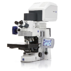
Confocal laser microscope, LSM700 Zeiss:

For conventional confocal analysis with three laser lines for fixed or live samples, the behavior of intra- and extracellular molecules in living cells can be studied by means of localized photobleaching (FRAP and FLIP), photoactivation or color preservation of fluorophores. It has 488, 550 and 639 nm laser lines.

For materials science analysis, this microscope allows the observation of morphology or defects in solids, such as microelectronic components, polymers, resins, deposits, minerals, ceramics, metals, etc.

Spectral multiphoton microscope, LSM780 Zeiss:

This technology allows for confocal spectral analysis; it has 405, 458, 488, 514, 561 and 633 nm laser lines. It has a polychromatic detector that allows the simultaneous detection of up to 10 markers, and its spectral detection system is capable of separating spectra, no matter how overlapping they are. It features a multiphoton pulsed laser adjustable from 680 to 1080 nm in 1-nm steps and uses low-energy (infrared) photons in extremely short pulses, making them less toxic to the biological sample.

This microscope can also generate deep three-dimensional images of living tissues and structures up to 0.5 nm thick with subcellular resolution. It is very useful in the study of subcellular structures, such as spines and axonal processes in neurons, that demand an image with a high signal-to-noise ratio over a large area or volume due to the wide distribution of axonal processes.

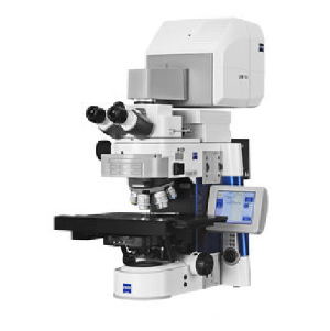
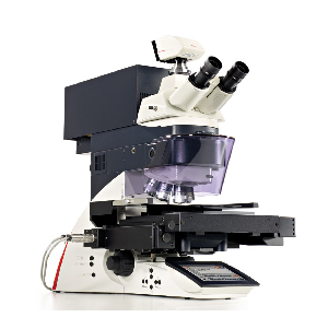
ELYRA - SIM Zeiss super-resolution laser microscope:

The resolving power of the light microscope is limited to approximately 200 nm in the lateral (xy) axis and 500 nm in the axial (z) direction due to the light diffraction limit. A technology with resolving power that exceeds the diffraction limit is structured light super-resolution (SIM). SIM allows the optical separation of molecules with a resolution twice as good compared that from conventional confocal microscopes. This technique has great application for example, in the study of protein architecture and dynamics in subcellular compartments. It also uses traditional fluorophores and has the 488 nm and 561 nm laser lines.

FT-IR Spectrum Frontier / Spotlight 400 Microscopy System, Perkin Elmer:

Using infrared light and Fourier transformation, this system generates spectral analyses of samples as small as 10 µm, and is ideal for materials, pharmaceutical, forestry, forensic, biomedical and biomaterials analyses.

FT-IR Spectrum Frontier / Spotlight 400 Microscopy System, Perkin Elmer:

Using infrared light and Fourier transformation, this system generates spectral analyses of samples as small as 10 µm, and is ideal for materials, pharmaceutical, forestry, forensic, biomedical and biomaterials analyses.

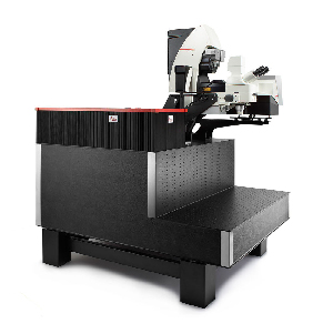
LMD 7000 Laser Microdissection Microscopy System, Leica:

This technique is used to dissect samples using a powerful UV laser, a process that is carried out through a precise optical system free of mechanical movements. Its high-power laser cuts both soft and hard samples. Histological samples can be cut, fixed, or stained. In addition, living cells, organelles, chromosomes, plant material and forensic materials can be extracted. The obtained material can be used for multiple techniques of cell biology, molecular biology and biochemistry, such as mutation analysis, SNPs, fingerprinting, LOH, FISH, RNA, gene expression analysis, microarrays, 2D PAGE, SELDI-TOF, MALDI-TOF , Western blotting, Dot Blot, cell cultures, tissue cultures, etc.

Cell Sorter BD FACSAria III Flow Cytometer:

Flow cytometry allows the simultaneous analysis and characterization of particles and / or cells and their subpopulations. The BD FACSAria III cell sorting equipment also allows the simultaneous separation of up to four populations, with a size range from 0.2 to 50 µm. The optical system is made up of two lasers, 488 and 633 nm, having the ability to detect eight colors and 10 parameters (including FCS and SSC). The equipment configuration is 5-3 (5 colors that are excited by the blue laser and 3 by the red) with the following filters:  Blue laser (488 nm): 780/60 BP, 695/40 BP, 585/42, 530/30.  Red laser (633 nm): 780/60 BP, 730/45 BP, 660/20 BP. It has integrated nozzles of 70, 85, and 100 µm, which allow us to analyze and separate most applications.

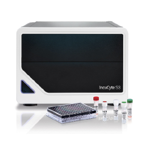
INCUCYTE S3 Live-Cell Analysis System:

This system allows for continuous monitoring of cells in culture by means of their visualization in real time using phase contrast microscopy coupled to detection of green and red fluorescence. HD fluorescence images are generated, while the cells remain stationary and undisturbed within the incubator. This system allows continuous analysis 24 hours a day for days or even weeks at a time, ensuring that no important cellular response or interaction is lost, performing an analysis based on images that provide both spatial and kinetic information. In addition, it has image processing tools that automate data analysis and help gain deeper insights from experiments. Continuous monitoring allows us to study cellular processes related to cellular health, including proliferation, apoptosis, cytotoxicity, and neurite growth; migration and invasion (chemotaxis, scratch wound), among others.

The equipment has capacity for up to six microplates at a time, in formats of up to 384 wells, allowing multiple experiments and applications to be programmed in parallel, leading to rapid optimization of variables and experimental conditions.

Scanning Electron Microscope VEGA3 EASYPROBE SBU (TESCAN)

This equipment is capable of offering a wide variety of information from the surface of the sample, but with higher resolution and with a greater energy range (up to 3 nm at 30 kV) than a typical SEM. As in SEM, an electron beam is swept over the surface of the sample, while the information that interests us is displayed on a monitor depending on the available detectors. This microscope features retractable SE and BSE detectors as well as an X-ray dispersive energy microanalyzer (EDS), which provides elemental semi-qualitative and semi-quantitative results from the live image. It has a variable pressure system, which makes it possible to work with high and low vacuum modes, allowing the analysis of non-conductive samples in their natural state without coating. It has a chamber with an internal diameter of 230 mm and a slit opening of 148 mm.

Very useful for characterizing materials such as metals, polymers, ceramics, composites, etc. Having applications in mineralogy, geology, paleontology, archeology, electronic engineering, chemistry, particle analysis, nanotechnology, botany, parasitology, pharmaceuticals, dental implants, forensics, etc.

Microscopio Electrónico De Barrido De Emisión De Campo VEGA3 EASYPROBE SBU (TESCAN)

El microscopio electrónico de barrido de emisión de campo (FESEM), es un instrumento que al igual que el SEM es capaz de ofrecer una amplia variedad de información procedente de la superficie de la muestra, pero con mayor resolución y con un rango de energía mucho mayor (hasta 3 nm a 30 kV). Al igual que en SEM se barre un haz de electrones sobre la superficie de la muestra mientras que en un monitor se visualiza la información que nos interesa en función de los detectores disponibles. Este microscopio cuenta con detectores SE y BSE retráctil además de un microanalizador de energía dispersiva de rayos X (EDS), el cual proporciona resultados elementales cualitativos y cuantitativos a partir de la imagen en vivo. Cuenta con un sistema de presión variable, lo que posibilita trabajar con modos de alto y bajo vacío permitiendo el análisis de muestras no conductoras en su estado natural sin recubrimiento. Posee una cámara con diámetro interior de 230 mm y abertura de porta de 148 mm.

Very useful for characterizing materials such as metals, polymers, ceramics, composites, etc. Having applications in mineralogy, geology, paleontology, archeology, electronic engineering, chemistry, particle analysis, nanotechnology, botany, parasitology, pharmaceuticals, dental implants, forensics, etc.

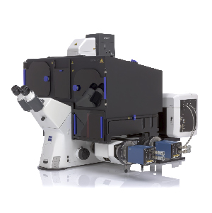
High resolution confocal spatio-temporal SP8 LIGHTNING, LEICA.

The latest acquisition of the CMA BIOBIO. It is a confocal equipment with software super resolution (LIGHTNING) and 8 kHz resonant scanner, which allows acquisition of up to 30 fps at 512x512, ideal for monitoring highly dynamic cellular processes, at a resolution of up to 130 nm lateral Microscope equipped with 4 laser lines (405, 488, 561 and 633 nm) and 4 spectral detectors, of which 3 are highly sensitive hybrid detectors (HyD) and the rest is a PMT for traditional applications. It has a 5% CO2 and temperature maintenance system for in vivo experiments and various objective lenses from 10 to 63x magnification, for most applications in biological sciences and medicine.


en_US